Mayo Clinic cardiac MRI research program seeks to improve access to diagnostic imaging
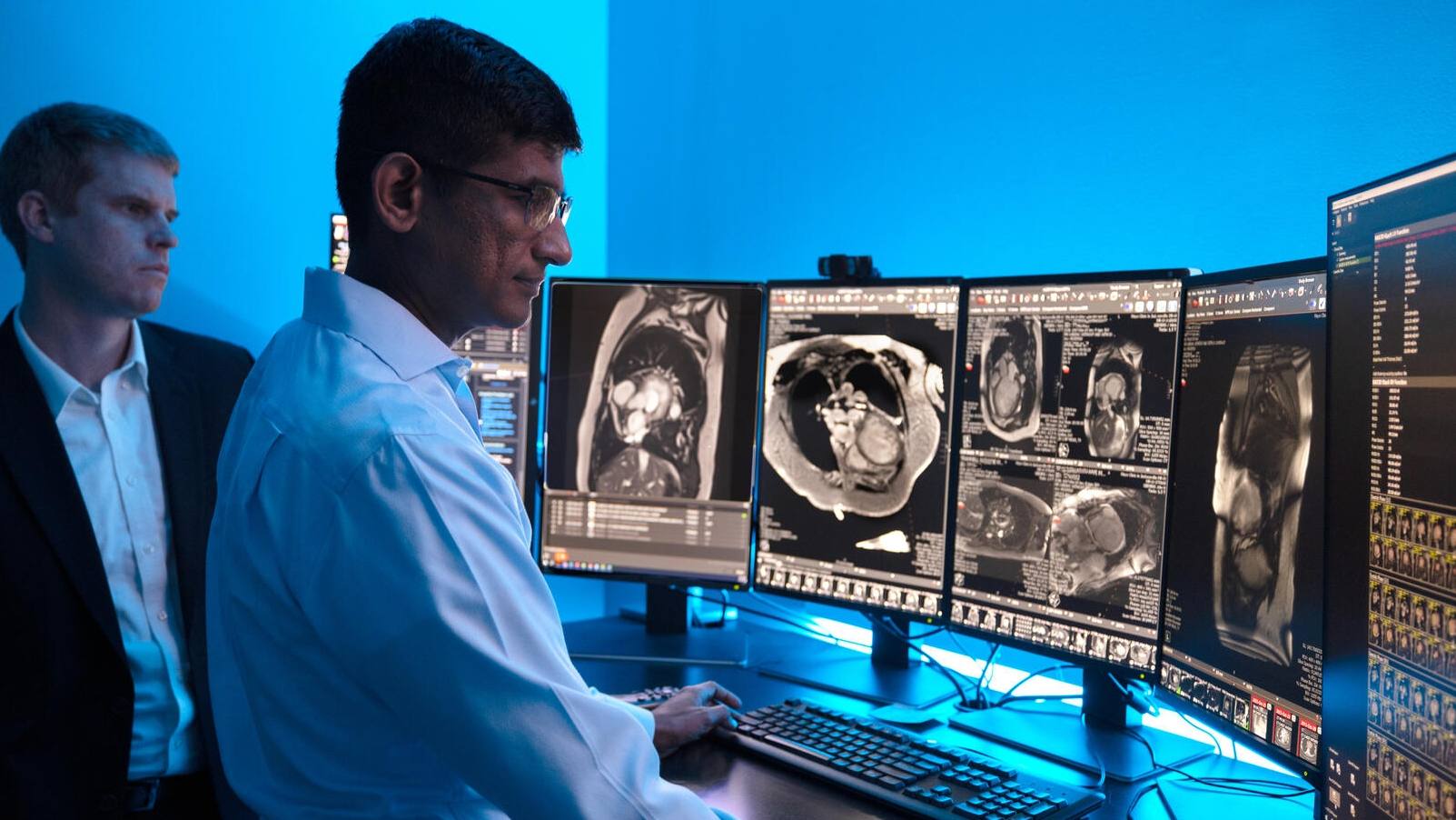

Just 5%–10% of people who need cardiac MRI get the exam because of barriers like cost, time and access, says Dr. Tim Leiner, a Mayo Clinic radiologist who, along with Dr. Jacinta Browne, a Mayo Clinic radiology researcher, leads the Artificial Intelligence for Cardiovascular Imaging Research and Exploration Program.
Imaging of the heart, especially with MRI, is an important part of diagnosing heart disease, the leading cause of death worldwide.
“That means they are operating without the best possible information. At Mayo the problem is lessened because we have so many resources, but, in general, this is a huge problem,” Dr. Leiner says.
Mayo Clinic, in collaboration with Philips, an international health technology company, is testing a low-field MRI scanner enhanced with artificial intelligence (AI) to shorten scan times and improve image quality.
The team’s work initially is searching for solutions in cardiac imaging while also looking at other parts of the body where their work can make an impact. They also hope to see cardiac MRI accessible to more people.
New ways of seeing

“Mayo Clinic is always investing resources and being at the forefront, trying to solve problems,” Dr. Leiner says. “I think the secret is to stay curious and surround yourself with people who are smarter than you. And you create a safe atmosphere where people can speak up and bring ideas to the table for an open discussion.”
The program’s approach seeks to blend hardware and software developments to achieve results the researchers hope will be as good as or better than current methods.
Dr. Leiner says the team wants to learn if a new type of scanner, a 0.6-Tesla (0.6T) low-field-strength MRI system, is able to produce high-quality images faster and more easily for patients.
“Radiology is about getting the piece of information from the imaging test that allows you to make the best decision in the patient’s care management, and once you have that information, you can move on,” Dr. Leiner says. “I think what we’re learning is that this lower field strength is entirely capable of giving you that information.”
Team of experts
The Artificial Intelligence for Cardiovascular Imaging Research and Exploration Program brings together Mayo Clinic physicians, physicists and scientists from across specialties, including Radiology and Cardiovascular Medicine, as well as Philips, which installed the research-prototype MRI scanner in One Discovery Square on the Mayo Clinic campus in Rochester, Minnesota. Philips is a collaborator on research with the system.
“It’s a truly multidisciplinary lab that I think can bring about a lot of progress because you’re getting viewpoints from scientists, clinicians and engineers,” Dr. Browne says. “That means that what you’re doing will be impactful for patient care.”

Dr. Browne says Philips’ presence on site enhances their research as well. “What is really great is the close collaboration that we have with Philips, the fact that we can just walk into each other’s offices and chat. And because we have an agreement with them, we have first access to a lot of the new work-in-progress products, and we’re at the table with them so we can also influence slightly the way in which they are going to go with some of those products,” she says.
“Mayo Clinic’s cardiac MRI research program has the potential to improve patient access to vital diagnostic imaging,” said Dr. Ioannis Panagiotelis, business leader of MR at Philips. “Their commitment to improving patient care through faster, more accessible, and personalized imaging aligns closely with our mission, and collaborating with their multidisciplinary team allows us to contribute technology that could make a meaningful difference for patients.”
The Artificial Intelligence for Cardiovascular Imaging Research and Exploration Program is looking at what improvements may be feasible for cardiac MRI in several areas:
- Is there a way to more uniquely tailor exams in real time?
- Can scan duration be reduced?
- Can imaging distortions created by medical implants be reduced?
- Can diagnosis be improved?
“We are investigating what we can do with such a system for cardiac imaging and for other body areas,” Dr. Leiner says. Both Mayo and Philips are contributing AI algorithms to integrate into the scanner.
Imaging the individual
“We’re looking at having a personalized cardiac MRI examination, which is going to be autonomous,” Dr. Browne says.
She adds that the goal is for images to be segmented and measured during the first scan sequence so it can be determined if the heart is functioning normally or if there are signs of cardiac disease. Those findings can then help determine — in real time — whether additional imaging is needed and, if so, what sequences will provide the necessary information to assist in treatment planning.
“You’re tailoring that examination, but you’re also potentially shortening the majority of the examination, so we’ll get some time savings,” she says, adding that the immediacy of the information would allow for additional imaging at that time to save the patient from having to return for a follow-up.
Faster scan time
Need for cardiac imaging exceeds scanner availability, Dr. Leiner says, so it’s important to use existing machines as efficiently as possible to ensure that patients get the scans they need when they need them.
He says the program is looking for ways to develop faster imaging protocols to scan patients quickly while getting the necessary clinical information for diagnosis and treatment planning. Cardiac exams are typically an hour long.
“One way we’re doing that is by using AI tools to shorten the image acquisition time, though if you shorten the acquisition time, you get lower signal-to-noise in MRI (meaning the images may not be as clear),” Dr. Leiner says. “But if you denoise the images that you get, those sort of lower-quality images, you can then reconstruct images that look as good as if they were acquired with a normal spatial resolution and machine settings.”
If successful, these tools may reduce scan time, he adds. “It’s about making this whole pipeline more efficient and more robust.”
Clearer images
With stronger MRI systems, most often 1.5T or 3T scanners, imaging patients who have implanted medical devices such as pacemakers can be challenging, Dr. Leiner says. With the higher-field-strength machines, those devices create distortions, called artifacts, in the images that can reduce anatomical visibility for physicians.
He says the researchers hope that the 0.6T scanner and AI solutions will help reduce the noise and improve the clarity in those images.
“What we need to do is come up with new ways in which we can get a higher signal, whether that is through the physics engineering of the pulse sequences we use in order to get the examination or whether it is through the use of AI, using denoising and image-sharpening techniques,” Dr. Browne says. “There are so many unanswered questions because we don’t know how this technology is going to work for all of the standard cardiac examinations.”
“It’s not inconceivable that in the future, AI algorithms will be able to reduce some artifacts at higher field strengths that you get in patients with devices or with other issues,” Dr. Leiner says.
Shared knowledge
Research from the Artificial Intelligence for Cardiovascular Imaging Research and Exploration Program will be shared during the Radiological Society of North America (RSNA) 111th Scientific Assembly and Annual Meeting Nov. 30—Dec. 4 in Chicago.
###
About Mayo Clinic
Mayo Clinic is a nonprofit organization committed to innovation in clinical practice, education and research, and providing compassion, expertise and answers to everyone who needs healing. Visit the Mayo Clinic News Network for additional Mayo Clinic news.
Media contact:
- Ethan Grove, Mayo Clinic Radiology, newsbureau@mayo.edu
The post Mayo Clinic cardiac MRI research program seeks to improve access to diagnostic imaging appeared first on Mayo Clinic News Network.